NIH Chest X-Ray Dataset
About
Chest X-ray exams are one of the most frequent and cost-effective medical imaging examinations available. However, clinical diagnosis of a chest X-ray can be challenging and sometimes more difficult than diagnosis via chest CT imaging. The lack of large publicly available datasets with annotations means it is still very difficult, if not impossible, to achieve clinically relevant computer-aided detection and diagnosis (CAD) in real world medical sites with chest X-rays. One major hurdle in creating large X-ray image datasets is the lack resources for labeling so many images. Prior to the release of this dataset, Openi was the largest publicly available source of chest X-ray images with 4,143 images available.
This NIH Chest X-ray Dataset is comprised of 112,120 X-ray images with disease labels from 30,805 unique patients. To create these labels, the authors used Natural Language Processing to text-mine disease classifications from the associated radiological reports. The labels are expected to be >90% accurate and suitable for weakly-supervised learning. The original radiology reports are not publicly available but you can find more details on the labeling process in this Open Access paper: "ChestX-ray8: Hospital-scale Chest X-ray Database and Benchmarks on Weakly-Supervised Classification and Localization of Common Thorax Diseases." (Wang et al.)
Data Limitations
- The image labels are NLP extracted so there could be some erroneous labels but the NLP labeling accuracy is estimated to be >90%.
- Very limited numbers of disease region bounding boxes (See BBoxlist2017.csv)
- Chest x-ray radiology reports are not anticipated to be publicly shared. Parties who use this public dataset are encouraged to share their “updated” image labels and/or new bounding boxes in their own studied later, maybe through manual annotation
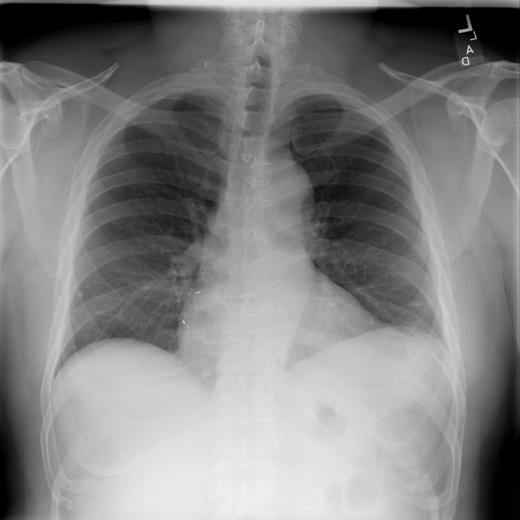
Sample Image